Project manager: Prof. Dr. Irinel Popescu
Formator: Dr. Florin Botea
Responsible of implementation: Dr. Mihail Pautov
Nr. of participants: 12
The workshop was divided into two days
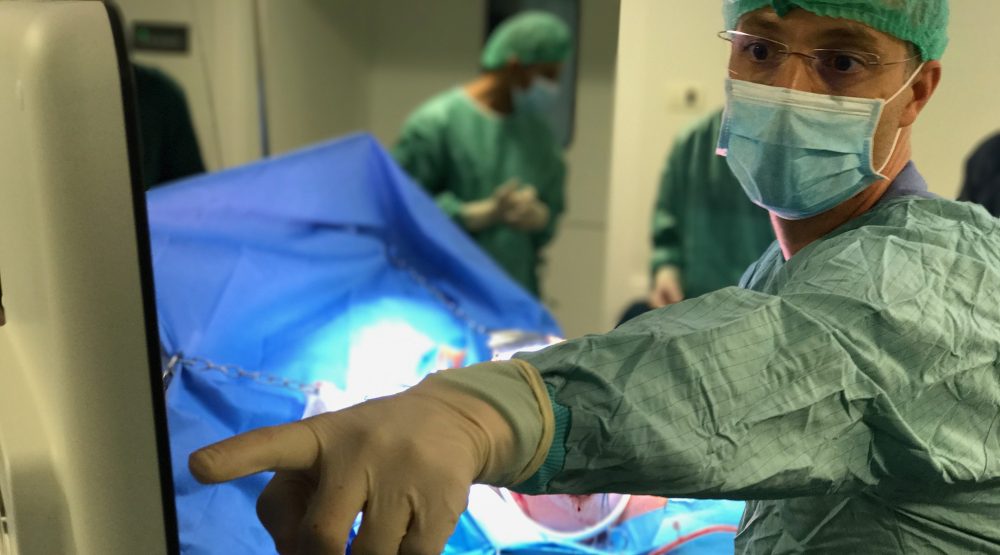
The first day, April 21, consisted of theoretical presentations with notions of liver anatomy, ultrasound, and principles of intraoperative liver ultrasound: indications, methods, ultrasound of the liver, and particular variants. After the theoretical part, the participants had access to the surgical room where they attended the intraoperative liver ultrasound in two interesting surgical cases: the first case, a 55-year-old patient with a hepatic tumor of about 7-8cm in the 8th hepatic segment. The participants had the opportunity to sonographically inspect the whole liver for other lesions, to characterize the lesion and to establish its relationship with the rest of the anatomical structures to establish a resection plan. The second case, a 59-year-old patient with a volumetric right hemorrhagic tumor (HCC) with percutaneous portal vein embolization one month ago in order to increase the post-embolization liver mass. The participants had the opportunity to inspect the left ventricle and establish the relationship between the tumor and Vena Haptica Media in order to establish the resection plan.
The next day: hands on procedures. The participants had access to the Experimental Surgery Hall of the Center for Excellence in Translational Medicine, where, under the guidance of the trainers, they were able to perform an intraoperative liver ultrasound on two large animals (pigs) with two dedicated ultrasound machines. Here the theoretical and practical notions accumulated on the previous day were strengthened, at the end of the workshop, each of the participants succeeded in the technique of intraoperative ultrasound, the intraoperative ultrasound inspection of the liver, the evaluation of the vascular anatomy and the detection of the anatomical variants, the diagnosis in the focal liver lesions and the surgical strategy according to this.
